bed bug bites on dark skin rarely present as the bright red welts shown in stock photos. Melanin-rich skin often masks erythema, producing instead subtle dark spots, purplish-brown discoloration, or small raised bumps that blend with surrounding tissue. Recognizing these atypical patterns matters because delayed identification allows infestations to establish while victims chase dermatological misdiagnoses.
Why Melanin Changes What You See
In my field work across diverse housing stock, I have watched residents with darker skin tones dismiss early signs for weeks. The inflammatory response that produces obvious redness in fair skin often manifests as dusky hyperpigmentation or barely perceptible swelling in phototypes IV through VI on the Fitzpatrick scale. Blood vessels dilate beneath the surface, but the overlying melanin absorbs and scatters the visible red wavelengths.
What remains visible tends to emerge hours or days after feeding. You may notice small, firm papules that feel slightly raised to the touch but appear only marginally darker than baseline skin tone. In some cases, the bite site develops a violaceous or deep brown hue that resembles bruising. This delayed color change often leads people to attribute marks to minor trauma rather than insect feeding.
The clustering pattern becomes your most reliable diagnostic clue. Bed bugs typically feed multiple times during a single encounter, creating linear or triangular groupings colloquially called "breakfast, lunch, and dinner." On dark skin, these clusters appear as subtle constellations of slightly darker dots—easy to miss under artificial lighting, more visible under natural daylight angled across the skin surface.
Post-Inflammatory Hyperpigmentation: The Lingering Tell
What I tell clients to monitor is not the acute reaction but the aftermath. Dark skin demonstrates heightened susceptibility to post-inflammatory hyperpigmentation (PIH), where melanocytes deposit excess pigment following any inflammatory insult. A bite that resolves in days on fair skin may leave a persistent dark macule for months on pigmented skin.
These residual marks create diagnostic confusion. They resemble solar lentigines, friction hyperpigmentation from clothing seams, or even early melanocytic lesions. The key distinguishing feature lies in distribution: PIH from bed bug bites clusters in exposed areas—arms, shoulders, neck, face—corresponding to skin left uncovered during sleep. Marks appear in crops that wax and wane with continued exposure, unlike static benign lesions.
I often see patients who have cycled through multiple topical prescriptions for presumed dermatitis while the actual infestation proliferated in their mattress seams. The PIH itself requires no treatment beyond sun protection to prevent darkening, but recognizing its origin prevents unnecessary medical expenditure and delayed pest control.
Visual Patterns Specific to Darker Skin Phototypes
Examination technique matters more than on fair skin. Raking light across the surface reveals subtle topography—minute elevations with central puncta that direct lighting obscures. Palpation frequently detects induration invisible to casual inspection.
| Fair Skin Presentation | Dark Skin Presentation | |
|---|---|---|
| Acute color | Bright erythema, pink to red | Dusky, violaceous, or brownish; may match baseline |
| Border definition | Sharp, inflamed edges | Poorly demarcated, blends gradually |
| Swelling visibility | Obvious wheal formation | Detected by touch rather than sight |
| Residual mark | Faint pink macule | Persistent hyperpigmentation, months duration |
| Cluster recognition | Visually obvious | Requires deliberate pattern search |
The central punctum—where mouthparts penetrated—often appears as a pinpoint dark speck rather than a red dot. This feature distinguishes bed bug bites from papular urticaria or other arthropod reactions, though it requires magnification or optimal lighting to appreciate on pigmented skin.
Differential Diagnosis: What Else Looks Similar
Several conditions mimic bed bug bites on dark skin, complicating self-diagnosis. Papular urticaria from flea bites produces similar clustered papules but typically concentrates on ankles and lower legs rather than upper body exposure zones. Mosquito bites generally present as isolated lesions with more pronounced surrounding edema.
Scabies burrows create linear or nodular patterns that can confuse inspection, though distribution favors finger webs, wrists, and genital areas rather than the exposed sleep surfaces bed bugs target. Dermatitis herpetiformis and other autoimmune blistering disorders produce grouped lesions but demonstrate chronicity and specific anatomical patterns inconsistent with intermittent nocturnal feeding.
More critically, bacterial folliculitis and early cellulitis can resemble inflamed bite reactions. Staphylococcal infections typically progress with increasing pain, warmth, and purulent drainage—features absent in uncomplicated bed bug bites. Any lesion that enlarges, becomes increasingly tender, or develops surrounding erythema spreading over hours warrants medical evaluation rather than continued observation.
Confirming the Source: Inspection Over Symptom-Chasing
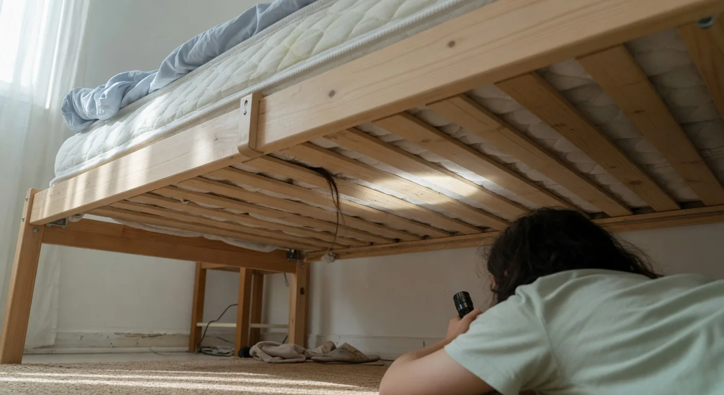
Relying on bite appearance alone courts misdiagnosis. What homeowners miss most is the necessity of finding harborage evidence. Bed bugs leave distinctive fecal spotting—small, dark, rust-colored deposits that smear reddish-brown when moistened—along mattress seams, box spring corners, headboard crevices, and nearby furniture joints.
Conduct inspection during daylight with a flashlight and credit card for crevice probing. Focus on the sleeping microenvironment within six feet of the bed. Shed skins, egg casings, and live insects confirm infestation regardless of skin findings. Eggs themselves measure approximately one millimeter, pearly white, and cemented to surfaces with adhesive secretion that resists casual dislodgement—washing alone rarely removes them, but sustained high heat in a clothes dryer typically terminates embryonic development.
I recommend photographing any skin findings and suspected harborage evidence contemporaneously. This documentation assists both dermatologists and pest management professionals in correlating clinical presentation with environmental confirmation.
When Medical Evaluation Becomes Necessary
Most bed bug bites resolve without intervention, but certain scenarios warrant professional assessment. Secondary bacterial infection from excoriation presents with increasing erythema, warmth, purulent drainage, or systemic symptoms like fever. Bullous reactions—large fluid-filled blisters—occur in some individuals and require wound management to prevent scarring, particularly consequential on visible skin.
Anaphylaxis, though rare with bed bugs, demands emergency care if systemic urticaria, respiratory compromise, or cardiovascular symptoms develop following exposure. Persistent pruritus disrupting sleep or extensive reactions across body surface area also justify dermatological consultation.
For residual hyperpigmentation, dermatologists may discuss options including topical retinoids or chemical exfoliants—discussions beyond pest management scope. The critical point: treat the infestation to stop new bites; treat the skin only for symptomatic relief or complication management.
Practical Steps for Dark-Skin Households
Prevention and early detection strategies require adaptation for populations where bite signs prove subtle. Encasements for mattress and box spring eliminate harborage and trap any resident bugs, providing visible evidence through the transparent membrane. Light-colored encasements specifically aid detection on dark bedding where fecal spotting might otherwise camouflage.
Regular laundering of bedding at temperatures exceeding 60°C (140°F), followed by complete drying cycles on high heat, addresses both mobile insects and heat-susceptible life stages. Vacuuming with crevice tool attachments removes debris and early-stage infestations before establishment.
When travel or used furniture acquisition introduces risk, immediate inspection of seams and joints prevents introduction. Quarantine protocols for secondhand items—encasement or heat treatment before bedroom integration—protect sleeping areas.
Reader Questions: Bite Appearance on Pigmented Skin
Can bed bug bites look completely flat on dark skin?
Yes. Some individuals, particularly those with prior sensitization, develop only macular hyperpigmentation without palpable elevation. These flat, dark patches often go unnoticed until clusters become apparent through pattern recognition.
Do bed bug bites always itch?
No. Pruritus varies dramatically by individual immune response. Some people experience intense nocturnal itching; others remain entirely asymptomatic despite repeated feeding. Absence of itching does not exclude infestation.
How long do dark marks from bites persist?
Post-inflammatory hyperpigmentation typically fades over weeks to months. Sun protection prevents darkening; persistent marks beyond six months warrant dermatological evaluation to exclude other pigmentation disorders.
Can the clustering pattern appear random rather than linear?
Bed bug feeding behavior produces varied configurations depending on fabric folds, skin contact points, and insect movement. While linear "breakfast, lunch, dinner" groupings are classic, scattered clusters and isolated lesions also occur. Pattern analysis supplements but never replaces environmental inspection.
Should I treat my skin before addressing the infestation?
Treating skin symptoms without eliminating the source guarantees continued exposure. New bites will appear as long as harborage persists. Prioritize inspection and professional pest management; address skin only for symptomatic complications.
Related guides — different questions
These articles cover overlapping topics but answer different questions. Pick the one that matches your situation.
- How to Stop Bed Bug Bites From Itching Naturally (Safe
Read if: Confirmed bites are itching and disrupting sleep tonight.
Frequently Asked Questions
What do bed bug bites look like on baby skin?
On babies, bed bug bites often appear as small red bumps or welts, sometimes in lines or clusters on exposed skin such as arms, neck, or face. They may look like hives or eczema, so check the crib area for bugs or spots on bedding.
Seek pediatric advice if bites swell rapidly, show infection, or your baby has breathing changes. Avoid strong OTC pesticides in nurseries without professional guidance.
What do bed bug bites typically look like and how can I tell them apart?
Bed bug bites often appear as small, red, itchy bumps, usually in lines or clusters, but reactions vary. Other insects or skin conditions can look similar, so it’s best to consult a healthcare provider for an accurate assessment.
- Common signs include raised welts, mild swelling, and itchiness.
- Bites may appear on exposed skin, especially around arms, neck, or face during sleep.
- Track new patterns and avoid scratching to lower infection risk.
Only a qualified professional can confirm bed bugs through inspection and evidence, not bite appearance alone.
What are the typical signs of bed bug bites on skin?
Bites often appear as small, red, itchy welts in a line or cluster. Reactions vary, so confirm with physical signs of bed bugs and professional guidance.
What does bed bug poop look like on walls?
Bed bug poop on walls looks like tiny dark dots or short streaks—often black, brown, or rust-colored. Spots are usually pinhead size and may smear when wiped with a damp cloth.
Check along baseboards, outlet covers, and behind headboards. A positive smear test (rusty streak) strongly suggests fecal stains, not ordinary dirt.